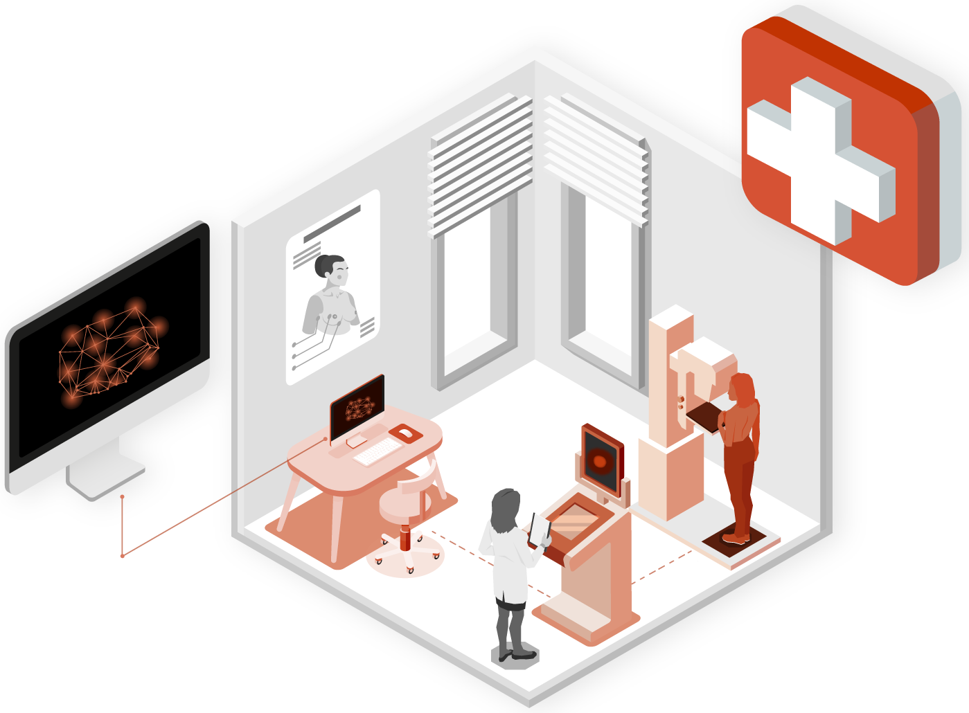
Algorithms for the reconstruction of the breast geometry from medical imaging

In collaboration with

Scenario

In the field of medical prevention, there are increasingly more technologies being developed to make analyses more effective and less invasive. One of the biggest challenges is that of ensuring the accuracy of the results so that they can support medical personnel in the diagnostic phase.
A biomedical equipment company has requested the support of Deix to improve the technology behind a device that they are developing, which is capable of providing support to medical personnel in diagnosing breast cancer
The challenge
The biggest challenge was to identify the region of interest for the analysis and reconstruct its 3D volume
The need was to improve the output of the detections of the new device by exploiting advanced algorithms to generate a result as representative of reality as possible.
The device that is the subject of our work bases its operation on mathematical models capable of identifying tissue properties from a set of optical and infrared detections. Deix played a crucial role in developing an algorithm capable of improving the reconstruction of breast geometry from the acquired images: the biggest challenge was to identify the region of interest for the analysis and reconstruct its 3D volume, eliminating the inherent noise of the acquisitions and considering the criticalities given by heterogeneous detections.
The solution
Deix has developed a robust algorithm that leverages various techniques of image analysis and computational geometry to combine acquired information and generate output.

The algorithm is able to accurately identify the region of interest for analysis, through a segmentation process that jointly uses 2D images and 3D acquisitions.

The use of both sources of data allows to overcome individual limitations of each (noise and imprecision of 3D data and poorly illuminated areas in 2D images) by reconstructing the 3D surface of the region of interest.

Techniques of image rasterization were used to map the information derived from the 2D image to the 3D model.
Results achieved
Week after week, the joint R&D work with the client has allowed for an increase in the accuracy of breast reconstruction, enabling the machine to provide increasingly effective support for the doctor's analysis.
The project has therefore achieved the agreed-upon objective, improving the output generated by acquisitions made by the device. One of the most interesting aspects was certainly working in incremental phases, refining the tool step by step, thanks to continuous feedback and experimentation. This is a typical approach for algorithm development projects, which proceeds through incremental testing in order to achieve the best possible result.
Future developments
The work methodology and algorithms used can certainly be utilized and replicated in different contexts: from industry to civil engineering, from production to security.
In particular, all areas - not just medical ones - where it is necessary to retrieve information from image acquisition, lend themselves to the implementation of algorithms and projects that use the same technologies and skills, even if for different purposes.
If you think you need our support for innovative activities of this caliber, contact us to discuss your needs with us
